Gray Matter Heterotopia


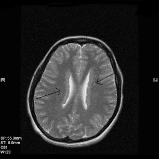
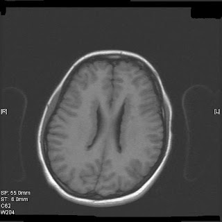
Gray matter heterotopia are common malformations of cortical development. From a clinical perspective, affected patients are best divided into three groups: subependymal, subcortical, and band heterotopia (also called double cortex). Symptomatic women with subependymal heterotopia typically present with partial epilepsy during the second decade of life; development and neurologic examinations up to that point are typically normal. Symptoms in men with subependymal heterotopia vary, depending on whether they have the X-linked or autosomal form. Nearly all affected patients that come to medical attention have epilepsy, with partial complex and atypical absence epilepsy being the most common syndromes.
Reference and detailed review in Neurology 2000;55:1603-1608 by Barkovich and Kuzniecky.
Consultant Radiologist ,VIMHANS and CEO-Teleradiology Providers
Editor-in-chief, The Internet Journal of Radiology
Director, DAMS (Delhi Academy of Medical Sciences)
 Reviewed by Sumer Sethi
on
Wednesday, May 07, 2008
Rating:


No comments:
Post a Comment