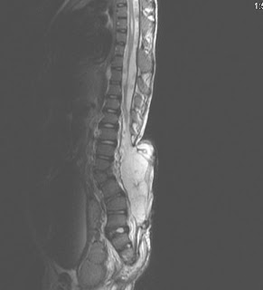
Chiari Malformation-MRI


MRI images showng lumbosacral myelomeningocele, dorsal syringohydromyelia and tonsillar herniation, classical Chiari II malformation.
Dr.Sumer K Sethi, MD
Consultant Radiologist ,VIMHANS and CEO-Teleradiology Providers
Editor-in-chief, The Internet Journal of Radiology
Director, DAMS (Delhi Academy of Medical Sciences
Chiari Malformation-MRI
 Reviewed by Sumer Sethi
on
Wednesday, June 18, 2008
Rating:
 Reviewed by Sumer Sethi
on
Wednesday, June 18, 2008
Rating:


No comments:
Post a Comment