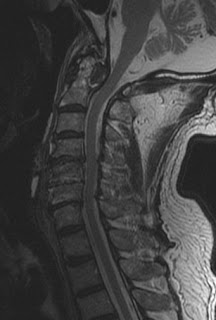
Craniovertebral Gout


This is an old male patient who is a known case of gout with raised uric acid levels and neck pain. MRI cervical spinhe revealed C1-2 erosions and marrow edema, with retro-odontoid soft tissue which is isointense on T1 WI and hypointense on T2 WI. There was no significant post gadolinium enhancement. CT scan revealed erosive disease and retro-odontoid high density soft tissue. There was no evidence of tuberculosis and rheumatoid factor repeatedly was negative. Although rare but still possible gouty cystal deposition was the provisional diagnosis made and patient was followed up after 3 months with no serial change.
Dr.Sumer K Sethi, MD
Consultant Radiologist ,VIMHANS and CEO-Teleradiology Providers
Editor-in-chief, The Internet Journal of Radiology
Director, DAMS (Delhi Academy of Medical Sciences
 Reviewed by Sumer Sethi
on
Saturday, July 26, 2008
Rating:


No comments:
Post a Comment