Cryptococcal Meningitis-MRI


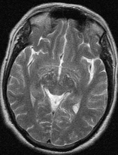
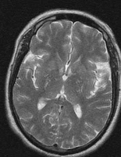
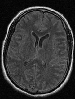
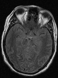
Cryptococcal organisms spread from the basal cisterns through the Virchow-Robin spaces to the basal ganglia, internal capsule, thalamus, and brainstem.The production of voluminous mucoid material may enlarge the perivascular spaces. MRI is more sensitive than CT scanning in demonstrating abnormalities such as dilated perivascular spaces. These manifest on T2-weighted MRIs as punctate, hyperintense, round or oval lesions that are usually smaller than 3 mm. This is 40 yr old man with altered sensorium clinically suspected meningitis. Cyptococcal infection was suggested and confirmed microbiologically.
Dr.Sumer K Sethi, MD
Sr Consultant Radiologist ,VIMHANS and CEO-Teleradiology Providers
Cryptococcal Meningitis-MRI
 Reviewed by Sumer Sethi
on
Wednesday, August 20, 2008
Rating:
 Reviewed by Sumer Sethi
on
Wednesday, August 20, 2008
Rating:


1 comment:
Radiological O****m.......
I see what u mean:)
Nice.....
Post a Comment