Radiology Case Discussion-Musculoskeletal Radiology


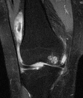
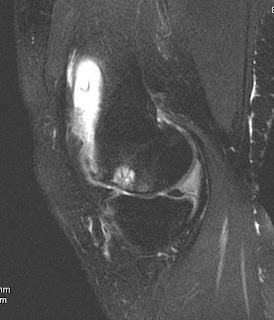
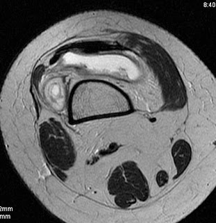
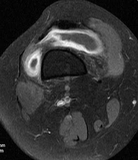
There is evidence of significant synovial collection in relation to the knee joint distending the supra-patellar bursa & medial / lateral recess. Synovium appears thickened & irregular. Fluid collection with septae is noted in relation to lateral aspect of femur with areas of signal suppression on GRE suggestive of haemosiderin staining. Areas of altered signal intensity appearing hyperintense on T2 fat sat images noted in patellar articular surface, medial condyle of femur & tibial plateau may indicate erosive process as disease history in chronic. Degenerative changes are seen in tibio-femoral & patello-femoral articulation in form of marginal osteophytes and articular cartilage thinning. On post gadolinium scans there is evidence of synovial thickening & enhancement. Nodular enhancement is noted in area noted in relation to lateral femoral condyle.
IMPRESSION: 32 yrs old female with synovial collection with synovial thickening & collection with septae in relation to lateral femoral condyle with haemosiderin staining, contrast enhancement and erosive process in patella, medial femoral condylar & tibial plateau, early degenerative changes. D/ D Includes synovial process like tuberculosis / RA / pigmented villo-nodular synovitis. ESR / RA factor is suggested. In view of haemosiderin staining PVNS is suggested as first differential. On follow up ESR and mantoux were done and were found to be negative.
Your comments and discussions are recommended.
Dr.Sumer K Sethi, MD
Sr Consultant Radiologist ,VIMHANS and CEO-Teleradiology Providers
Editor-in-chief, The Internet Journal of Radiology
Director, DAMS (Delhi Academy of Medical Sciences)
Sr Consultant Radiologist ,VIMHANS and CEO-Teleradiology Providers
Editor-in-chief, The Internet Journal of Radiology
Director, DAMS (Delhi Academy of Medical Sciences)
Radiology Case Discussion-Musculoskeletal Radiology
 Reviewed by Sumer Sethi
on
Wednesday, January 28, 2009
Rating:
 Reviewed by Sumer Sethi
on
Wednesday, January 28, 2009
Rating:


2 comments:
Informative! Thanks...
sir, i'm consultant radiologist in sikkim manipal institute of medical sciences, gangtok.
the subchondral hyperintense areas seen, are they secondary to degenerative process or primarily part of PVNS.
how will we differentiate from infective patholgy?
Post a Comment