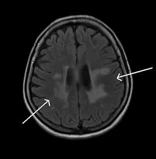
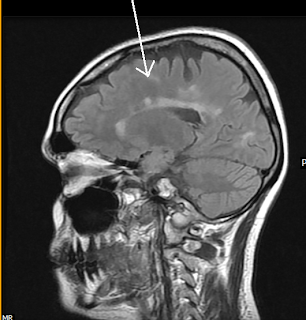
Multiple Sclerosis-MRI


Multiple discrete variable sized ovoid perpendicularly directed T2W and FLAIR hyperintense lesions (plaques), appearing iso-hypointense on T1W images and hyperintense on T2W images involving bilateral periventricular and subcortical white matter regions, including the calloso-septal interface. 35 year old female, case reported by Teleradiology Providers
Multiple Sclerosis-MRI
 Reviewed by Sumer Sethi
on
Monday, June 22, 2009
Rating:
 Reviewed by Sumer Sethi
on
Monday, June 22, 2009
Rating:


No comments:
Post a Comment