Spinal metastasis-MRI


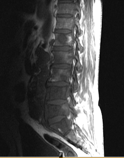
This is MRI lumbar spine of a 70 yr old male who came to us with complaints of back pain and pain in both lower limbs. It shows evidence of osseous destruction along with marorw signal abnormality of multifocal vertebral bodies involving all lumbar vertebral bodies, sacral ala. There is epidural soft tissue component with involvement of the posterior elements, appearing heterogeneously hyperintense on STIR and hypointense on T1W along with compromise of neural sac.
Discussion:
Four MR patterns of vertebral metastatic disease are seen – focal lytic, focal sclerotic, diffuse inhomogenous, diffuse homogenous. The most common among them is focal lytic lesions characterized by low signal intensity on T1 and high on T2. Pedicle destruction is more in favour of metastatic etiology. Pathologic compression fractures are also seen and show comparatively low signal intensity on T1 and high signal on T2 as compared to benign osteoporotic fractures which are mostly isointense on all sequences.
Case by Teleradiology providers
Spinal metastasis-MRI
 Reviewed by Sumer Sethi
on
Friday, November 06, 2009
Rating:
 Reviewed by Sumer Sethi
on
Friday, November 06, 2009
Rating:


3 comments:
Thanks, I like this post it is very good and informative. I am sure that this post will be very helpful.
A great article indeed and a very detailed, realistic and superb analysis of the current and past scenarios. I would like to thank the author of this article for contributing such a lovely and mind-opening article.
Thank you for this post, it helps greatly.
Post a Comment